
Note concerning the publication date of BfR2GO
BfR2GO 1/2026 was released in June 2026. The next issue will be published in December 2026. Internal Link:Subscribe
A ball of tissue
Listen to the article:
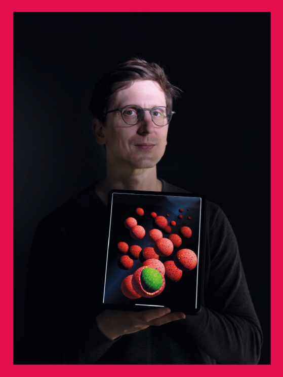
Biochemist Dr Julian Heuberger cultivates intestinal organoids, miniature replicas of organs. In this way he simulates the digestive system and wants to replace animal experiments.
At first glance, the object examined under the microscope gives the impression of being a small, glinting ball. Magnification reveals a layer of tightly packed rows of long cylindrical cells on the ball’s surface. Beneath these surface cells, the other cells appear fibrous and randomly arranged. All cells organised in this ball-like structure originate from the intestine. Its scientific term is “organoid.” It is a tiny, very similar version of a “real” organ. Organoids are the subject of intensive research and development because they pave the way for new scientific advances, including the use of organoids to help replace animal experiments.
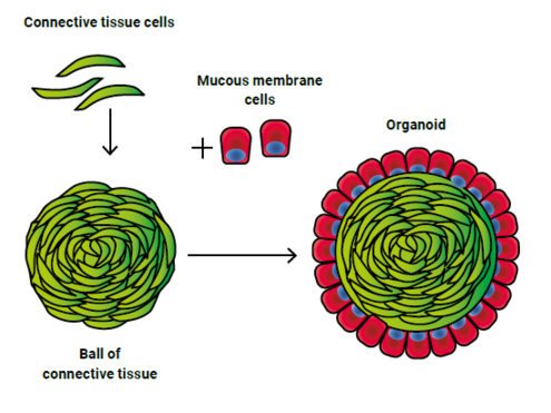
The man behind the ball-shaped organoids is Dr Julian Heuberger from the German Centre for the Protection of Laboratory Animals (Bf3R) at the German Federal Institute for Risk Assessment (BfRshort forGerman Federal Institute for Risk Assessment). Heuberger cultivates intestinal organoids from animal and human cells. The human intestinal organ consists of several layers. The singlelayered epithelium as the “lining” towards the inside. It forms the intestine’s mucous membrane which absorbs nutrients from the bolus. Underneath the epithelium is the connective tissue (stroma) interlaced with blood vessels, followed by an outer muscle layer.
Self-organising organoids
How can this be recreated in the laboratory? Heuberger has found an elegant solution. Heshort forhelium combines stroma cells from the intestine and the corresponding epithelial cells in a bioreactor – a special vessel for cultivating cells. Then what scientists call “self-organisation” occurs. The epithelial cells huddle up to the stroma cells, fuse with them and continue to mature. The organoid-ball organises itself. “After a few days the core of the connective tissue is completely covered with epithelium,” says Heuberger.
The colonic organoids produced in this way have significant advantages compared to earlier organoid models. The addition of growth-promotion factors is only required to a very limited extent since the stroma itself produces growth factors. In addition, these organoids can be composed with numerous macrophages (phagocytes that belong to the immune system).
The organoids thus have another essential element of the “real” intestine, as this is rich in cells for immune defence. This method of culturing organoids enables them to survive for weeks and be produced at a precisely defined, standard size. This significantly increases the range of applications for test systems.
Inside out
The greatest advantage of this culture technique, however, is the easily accessible epithelial surface of the organoid. The epithelial cells cover the stroma core like a thick lawn. This greatly facilitates the scientific study of the intestinal barrier, in order to, for example, better understand infections, metabolism and the influence of medications or chemicals on the organism. “We have turned the intestine inside out,” explains Heuberger. “In a sense, the epithelium is “bathing” in the test liquid, whereas in classic organoid cultures, the epithelial surface could only be reached with difficulty.”
A long road
![[Translate to Englisch:] Mikroskopaufnahme Organoid](/assets/_processed_/b/2/csm_ausgabe-1-2025-organoide_microcope_b6aa269297.jpg)
Matured colonic organoids
Even though the idea of artificial miniature organs is impressive and currently all the rage in the scientific community, the road to them being used as replacement for legally required animal experiments is still a long one. The intestine is just one part of the body, albeit an important one. Thus, intestinal organoids offer advantages such as the ability to test substances and make preselection before an animal experiment is necessary.
Cooperation with other laboratories is also important to the scientists as to date almost everyone has their own “recipe”. “This might be sufficient for academic questions,” says the researcher. “But it makes standardisation difficult, which is essential for the use of alternative methods to animal experiments.” To find joint solutions Heuberger cooperates with institutes such as the Berlin University Clinic Charité, the TU Berlin and the Max Delbrück Center for Molecular Medicine in Berlin-Buch.
After multiple scientific stops, Julian Heuberger has now found the ideal place at Bf3R to put his ideas into action. Both the scientific environment of the Bf3R and its proximity to the Berlin biomedical research institutes contribute to this. Heuberger has been at the Bf3R since 2023 – and it already appears to be a very organic relationship.